. 2018 Dec 14;7:e41050. doi: 10.7554/eLife.41050

Figure 5. Relative surface expression modulates mismatch coaggregation behavior.

(A) Western blot of biotinylated membranes showing low, medium, and high surface expressing populations of Pcdh1 after DNA titration. (B) Representative images of mismatch coaggregation assays mixing Pcdh1+Pcdh7 cells with Pcdh1 (low, medium, and high) cells. Scale bar, 100 μm. Results for each assay were determined from three independent electroporations. (C) Mean CoAg values show varying the expression levels in Pcdh1 cells did not alter the coaggregation behavior (interfacing), but did affect the degree of interfacing. Error bars indicate ±SEM. Dotted lines indicate thresholds for change in coaggregation category. (D) Representative images of mismatch coaggregation assays where the relative expression levels of co-expressed δ-Pcdhs were varied. Pcdh1High+Pcdh7Low cells and their complement, Pcdh1Low+Pcdh7High cells, were combined with cells expressing a given δ-Pcdh population. The two images in a given column (e.g. Pcdh1, column 1) illustrate the differences in coaggregation behavior when mixed with these two populations. (E) Mean CoAg values for (D), each bar indicates values for the top image in a given column vs. values for the lower image in a given column. Error bars indicate ±SEM, * indicates p≤0.05, **p≤0.01, ***p≤0.001. Results for each assay were determined from four independent electroporations.

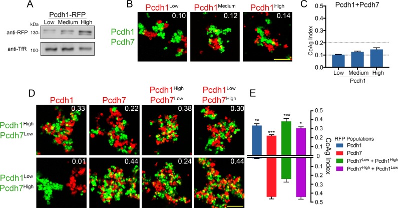
Figure 5.

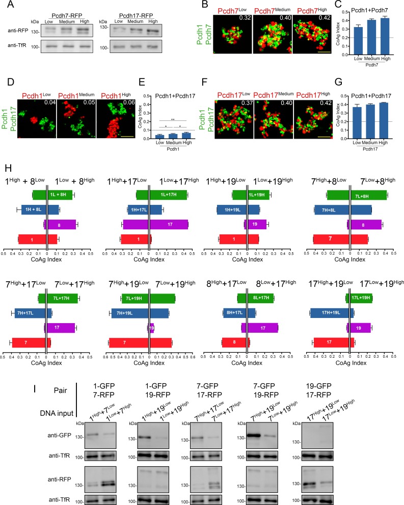
Figure 5—figure supplement 1. Effects of surface expression levels on mismatch coaggregation behavior.

Figure 5—figure supplement 1.

(A) Western blots of purified membranes showing low, medium, and high levels of Pcdh7 and Pcdh17 surface expression after DNA titration. (B,D,F) Representative images of mismatch coaggregation assays with (B) Pcdh1+Pcdh7 and (D,F) Pcdh1+Pcdh17 cells. (C,E,G) Mean CoAg values show varying surface expression in cells expressing a single δ-Pcdh did not fundamentally alter coaggregation behavior. Dotted lines indicate the threshold to change coaggregation categories. Cells still intermixed (Pcdh7, Pcdh17) or segregated (Pcdh1), although small changes in CoAg values do occur with increasing expression. Error bars indicate ±SEM, * indicates p≤0.05, **p≤0.01. Results for each assay were determined from three independent electroporations. (H) The impact of changing relative expression levels in cells expressing two δ-Pcdhs was tested for eight additional pairs of δ-Pcdhs. Each graph shows the mean CoAg values obtained for each mismatch assay. For example, the first graph shows the effects of varying relative expression of Pcdh1+Pcdh8. The left half of this graph shows results from assays using Pcdh1High+Pcdh8Low cells, while the right half shows results using the opposite conditions (Pcdh1Low + Pcdh8High). The impact of changing these relative expression levels is easiest to interpret by comparing the left and right half of each horizontal bar. For example, for the first bar (green), Pcdh1Low+Pcdh8High cells intermix more strongly with cells expressing Pcdh1Low+Pcdh8High than with Pcdh1High+Pcdh8Low cells. Pcdh8 cells (purple bar) intermixed with Pcdh1Low+Pcdh8High cells, but segregated from Pcdh1High+Pcdh8Low cells. Changing the relative level of expression in co-expressing cells can therefore change the category of coaggregation behavior. Error bars indicate ±SEM. Results for each assay were determined from two independent electroporations. (I) A subset of conditions shown in (H) and in Figure 5D were tested to confirm high/low DNA inputs resulted in changes in relative surface expression. Compare relative signal from the top half of each lane to the bottom. Note that the goal was to generate different expression ratios between the two lanes for each pair shown, and not to generate ‘high’ or ‘low’ surface expression.