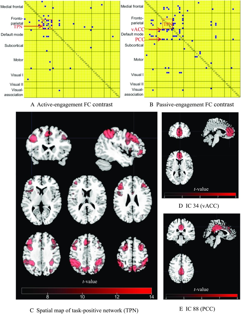
Figure 3. .
FC contrast maps between HE-FC and LE-FC during 2-back working memory task and spatial maps of ICs highlighted in two contrast maps. (A) Active-engagement FC contrast (HE > LE). Only links that were significant at a FDR-corrected p value of 0.01 were kept. The IC index is also displayed along the diagonal cell. The task-positive network (TPN) for working memory task (IC 64, 77, 78, 84, and 98) are highlighted by the rectangle. (B) Passive-engagement FC contrast (LE > HE). IC 34 and 88 pointed at by arrows are ventral anterior angular cortex (vACC) and PCC, respectively, which are more coupled to TPN during passive engagement. (C) A composite spatial map of task-positive ICs. (D) The spatial map of IC 34 (vACC). (E) The spatial map of IC 88 (PCC).

