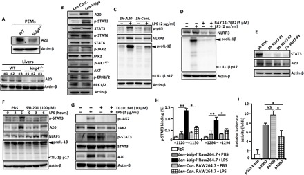
Fig. 2. VSIG4 inhibits the transcription of Nlrp3 and Il-1β via activating the AKT-STAT3-A20 axis.

PEMs and liver tissues were isolated from Vsig4−/− mice and their C57BL/6 WT littermates. (A) Western blot analysis of A20 (each number represents an individual mouse). RAW264.7 cells were transfected with Len-Cont. or Len-Vsig4. (B) Cell extracts were immunoblotted for the indicated protein. (C) A20 in RAW264.7 cells was silenced by shRNA, cells were further treated with LPS (2 μg/ml) for an additional 3 hours, and cell extracts were immunoblotted for the indicated protein. (D) PEMs were treated with LPS (2 μg/ml) in the presence of NF-κB inhibitor BAY 11-7082 (5 μM), and cell extracts were immunoblotted for the indicated molecules after 6 hours. (E) The transcription of Stat3 in RAW264.7 cells was silenced by shRNA, and cell extracts were immunoblotted for A20 and STAT3. LPS-primed RAW264.7 cells were treated with (F) the STAT3 inhibitor S3I-201 (100 mM) and (G) the JAK2 inhibitor TG101348 (10 μM), and cell extracts were immunoblotted for the indicated molecules after 12 hours. (H) ChIP-qPCR analysis of the enrichment of p-STAT3 binding to the potential binding sites in the A20 promoter region in Vsig4+RAW264.7 cells after 3 hours of LPS (2 μg/ml) administration. (I) Analysis of luciferase activity of A20 gene promoter constructs by luciferase reporter assay. Error bars indicate SEM. NS, not significant. *P < 0.05 and **P < 0.01 (Student’s t test). Data represent one out of three biological replicates, with three technical replicates each at least.
