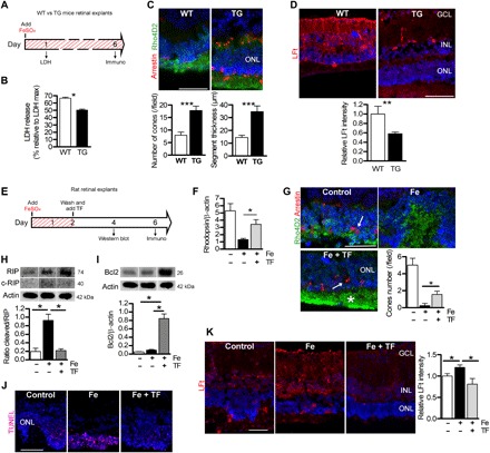
Fig. 3. Expression of TF protects retina explants exposed to iron.

(A) Illustration of timed workflow for retinal explants from WT and TG mice expressing hTF continuously exposed to 1 mM FeSO4. LDH release was measured after 1 day and immunostaining after 6 days. (B) LDH release was lower in culture medium from TG explanted mice compared to WT explants. Mann-Whitney test (n = 6), *P = 0.03. (C) Number of cones stained by arrestin (arrow) and length of rod segments stained by rhodopsin (Rho4D2) were quantified and were higher in TG explants. Mann-Whitney test (n = 3), ***P < 0.001. (D) Quantification of immunostaining intensity for markers of iron storage LFt was decreased in TG explants compared with WT explants. Mann-Whitney test (n = 3), **P < 0.01. (E) Illustration of timed workflow for TF treatment on iron-exposed explants: Adult rat retinas were exposed for 2 days with 1 mM FeSO4. Control explants were cultured with medium alone. Afterward, the medium was completely replaced and explants were treated with hTF (50 mg/ml) for 2 or 4 days. (F) Quantitative analysis of rhodopsin protein by Western blotting show increased rhodopsin protein expression in TF-treated iron-exposed explants (Fe + TF). Mann-Whitney test (n = 3), *P = 0.021. (G) Rhodopsin and arrestin immunostaining revealed protection of rod segments (Rho4D2, asterisk) and cones (arrow) by TF treatment (Fe + TF). Quantification of cone nuclei showed more cones in explants treated with TF than those exposed to FeSO4. Mann-Whitney test (n = 3), *P = 0.036. (H) Western blotting and quantitative analysis of RIP kinase demonstrated higher full form and cleaved form of the proteins in iron-exposed retinal explants. The cleaved form of RIP reported on RIP full form was reduced when TF was used to treat iron-exposed explants. Mann-Whitney test (n = 3), *P = 0.028. (I) Antiapoptotic Bcl2 protein, detected by Western blotting, was increased in TF-treated iron-exposed explants. Mann-Whitney test (n = 3), *P = 0.028. (J) TUNEL-positive cells in the ONL were reduced by TF treatment. (K) Immunostaining of iron storage marker ferritin light chain was significantly lower in explants treated with TF (Fe + TF) than without treatment (Fe). Fluorescence intensity was reported relative to control conditions. One-way analysis of variance (ANOVA), Bonferroni post test (n = 3), *P < 0.05. Scale bars, 100 μm. All values are represented as mean ± SEM.
