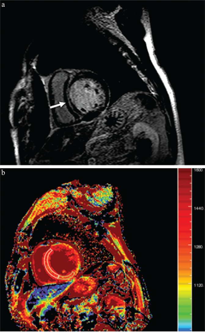
Fig. 4.

Imaging for a 60-year-old man with dilated cardiomyopathy and septal fibrosis. (a) Late gadolinium enhancement (LGE) was found in the mid-wall of the interventricular septum (arrow). (b) Non-contrast-enhanced T1 mapping shows that the native T1 value of the septal region including LGE (enclosed by a white line) is 1382.2 ms, which is more than 1349.4 ms and 1.2 standard deviation (SD) above that of the minimum T1 value (enclosed by a green line, 1262.4 ms; SD, 62.0 ms) in this patient.
