
Figure 2.
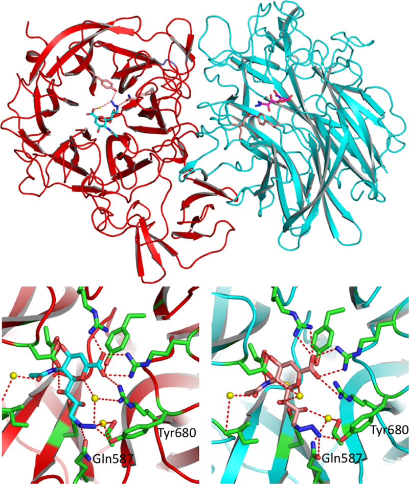
The novel complex structure between SpNanA and Neu5Ac9N32en (2). There are two subunits. A (red) and B (blue) in an asymmetric unit shown in the up panel. The bound Neu5Ac9N32en molecules in subunits A and B are shown in light blue and pink sticks, respectively. The close up views of Neu5Ac9N32en binding sites in subunits A (left) and B (right) are shown in the lower panel. The side chains of amino acid residues involving in the hydrogen bond interactions with Neu5Ac9N32en are shown in green sticks. Potential hydrogen bond interactions between the Neu5Ac9N32en and the protein are shown in red dashed lines.