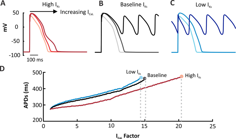
Figure 2. Altering the contribution of IKs within the same model influences arrhythmia susceptibility.

ICaL was progressively increased in each model to induce EADs and arrhythmic behavior. ICaL factor refers to the increase in channel permeability coefficient (1.0 equals control level).(A) APs simulated in the High IKs version of the O’Hara model at baseline and with ICaL augmentation (7.5 and 15.5 times). Plot shows the 92nd beat simulated at a 1 Hz pacing rate. (B) APs simulated in the baseline O’Hara model with the same ICaL perturbations. Plot shows the 91st beat. (C) Low IKs version simulated under the same conditions. Greater APD prolongation and increased number of EADs are produced in the low IKs version whereas the high model cells are protected from arrhythmogenic dynamics. Plot shows the 91st beat. (D) All three versions of the model were run under a wide range of ICaL factors and plotted against APD. The end of each line represents the last factor before an EAD forms The High IKs model requires a greater increase in the total ICaL current to induce an EAD.
