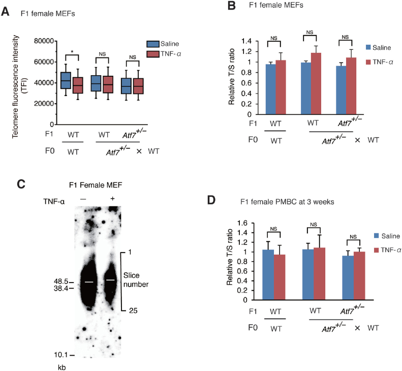
Figure 2.
Paternal TNF-α treatment does not induce telomere shortening in the female offspring. (A, B, C) Paternal TNF-α treatment did not induce telomere shortening in MEFs from female offspring. WT or Atf7+/– 8-week-old male mice (F0) (n = 3) were daily administered with TNF-α as described in Figure 1A, and then mated with WT female mice (n = 3). MEFs (n = 3 from three independent pregnant mice) were prepared from female offspring (F1 mice) for measurement of telomere length by Q-FISH (A). *, P < 0.05; NS, not significant. Raw data of Q-FISH are shown in Supplementary Figure S2. Telomere length of MEFs (n = 3, 5; 3, 4; 5, 3 for each group from three independent pregnant mice) were measured by Q-PCR (B). Relative telomere length, expressed as the T/S ratio, is shown ± SD. NS, not significant. Telomere length was also measured by telomere restriction fragment (TRF) assay using the G24 probe (C). Three primary MEFs from independent pregnant mice were used. The radioactivity of each slice in each lane (slice number is shown on the right) was quantitated using Image Analyzer, and the position of mean radioactivity was calculated. White bars indicate the position (slice number) of mean radioactivity in various samples. (D) Paternal TNF-α treatment did not induce telomere shortening in PMBCs from female offspring. Paternal mice were treated as above. PMBCs (n = 6, 3; 6, 6; 6, 5 for each group from three independent pregnant mice) were prepared from the 3-week-old female pups (F1), and telomere length was determined by Q-PCR as above. NS, not significant.

