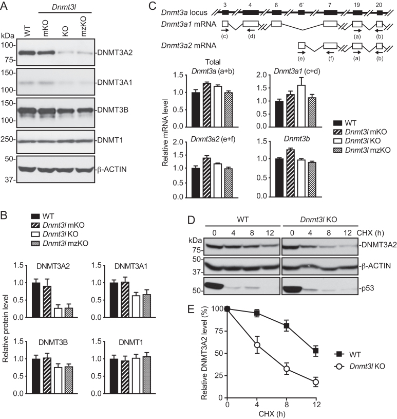
Figure 5.
DNMT3A is unstable in the absence of DNMT3L. (A and B) Western blot analysis of DNMT proteins in DNMT3L-deficient mESCs. Shown are representative blots (A) and quantification of the data (mean + SD from four independent experiments) by densitometry using ImageJ (B). (C) RT-qPCR analysis of Dnmt3a and Dnmt3b mRNAs in DNMT3L-deficient mESCs (mean + SD from two independent experiments). The Dnmt3a locus and the two major Dnmt3a transcripts, Dnmt3a1 and Dnmt3a2, as well as the locations of the primers (a–f), are schematically shown at the top. (D and E) Analysis of DNMT3A2 protein stability by inhibiting protein synthesis with cycloheximide (CHX) and then monitoring DNMT3A2 levels by Western blot for different periods of time. p53 was used as a positive control for the effect of CHX treatment, and β-ACTIN was used as a loading control. Shown are representative blots (D) and quantification of the data (mean ± SD from three independent experiments) by densitometry using ImageJ (E).

