Figure 2.
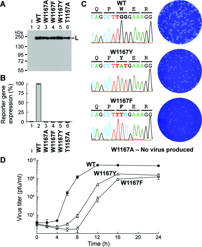
The W1167 residue in VSV L is essential for VSV gene expression and propagation in host cells. (A, B) The VSV mini-genome assay was performed with plasmids expressing N, P, and C-terminal FLAG-tagged L (WT or mutant). L proteins expressed in the transfected cells were detected by Western blotting with anti-FLAG antibody (A). The blot is a representative of three independent experiments. Lane 1 indicates no L plasmid. The graph shows relative expression levels of a reporter gene product (CAT), where expression levels in cells transfected without (column 1) and with (column 2) the WT L plasmid were set to 0 and 100%, respectively (B). The reporter gene product was not detected in lysates of cells expressing the indicated W1167 mutants as well as T1157A (negative control) (7) in the three independent experiments. (C) Effects of the indicated mutations on rVSV generation were examined. When rVSVs were viable, the mutation sites in rVSV genomes were sequenced (left). Plaque phenotypes of WT and mutant rVSVs were compared (right). (D) BHK-21 cells were infected with rVSV harboring the WT (closed circles), W1167Y (open triangles) or W1167F (open diamonds) L gene at a multiplicity of infection of five plaque-forming unit (pfu) per cell and cultured for 24 h. Single-step growth curves were obtained by titration of the rVSVs in the culture supernatants harvested at the indicated time points by a plaque assay. Symbols and error bars represent the means of titers and standard deviations, respectively (n = 3).

