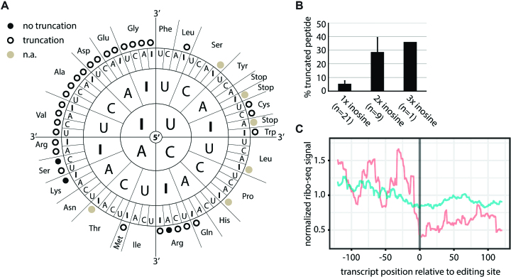
Figure 5.
Inosine causes ribosome stalling. (A) Black dots indicate exclusive detection of full-length peptide, circles indicate additional truncated peptides. Codons with inosine only in the wobble position or STOP codons were omitted (n.a.; gray dots). (B) Percentage of truncated peptide detected for different numbers of inosines in the codon are shown. Error bars = s.e.m. (C) Inosine levels at known editing sites were calculated from brain mRNA-seq data. Ribosome profiling data for sites showing editing (red) or no editing (blue) were normalized, weighted by editing rate, merged, and centered on the editing site. The coverage from position −125 to +125 relative to the editing site is given.

