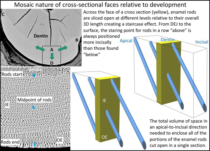
Figure 10.

Schematic illustration of the multidirectional developmental pattern for enamel and the step‐like arrangement of enamel rods created relative to the cross‐sectional plane of the mandibular mouse incisor. Low‐magnification BEI image of a cross‐section of the enamel layer covering the labial side of a mandibular mouse incisor (top left side). The boxed area is shown at higher magnification at the bottom. On the right side are schematic drawings illustrating how enamel rods project into or out of the plane of section to varying degrees depending upon their location across the thickness of the enamel layer. Enamel rod profiles, each representing a single 2D slice from the much larger 3D enamel rod, found near the DEJ are the most apical starting points for rods projecting mostly in an incisal direction from the plane of section, whereas rod profiles seen near the outer surface are cut at the incisal end of rods projecting in an apical direction backwards into the tissue block. Rod profiles seen in the middle of the enamel layer would have half their 3D length projecting apically and the other half projecting incisally, with all other profiles inbetween projecting predominately incisally (above midpoint) or apically (below midpoint). During development, enamel formation begins near the DEJ at the central side of the tooth (boxed area, location A) and spreads as a wave in a mesial direction (arrow to location B) and lateral direction (arrow to location C) as the enamel layer increases in thickness by appositional growth (arrow to location D) (Smith & Warshawsky, 1976). A cross‐section of the incisor therefore represents a composite image built up over time of the creation of lamellar sheets of rods stacked in a step‐like arrangement one behind the other with alternating tilts. This formative process spreads as a wave to the mesial and lateral sides of the labial surfaces so that location A begins its development before location B, followed by location C, thereby creating a time composite of development across the whole enamel layer. The thin ring of rod profiles abutting the DEJ and those abutting the outer surface define the entire volume of 3D enamel rod space sampled by the cross‐section.
