
Figure 4. Stimulation of α1ARs increases glutamatergic drive onto vPAGDA neurons in an AMPA receptor-dependent, action potential-independent manner.
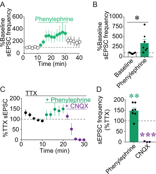
A-B) Bath application of phenylephrine (3 μM) significantly increased sEPSC frequency in vPAGDA neurons (n = 7; *p < 0.05). C) TTX (1 μM), phenylephrine (3 μM), and CNQX (10 μM) were cumulatively applied to slices and sEPSCs were recorded from vPAGDA neurons. D) Phenylephrine in the presence of TTX increased sEPSC frequency compared to sEPSCs during TTX alone (n = 7; **p < 0.01), and CNQX abolished sEPSCs (n = 3; ***p < 0.001).