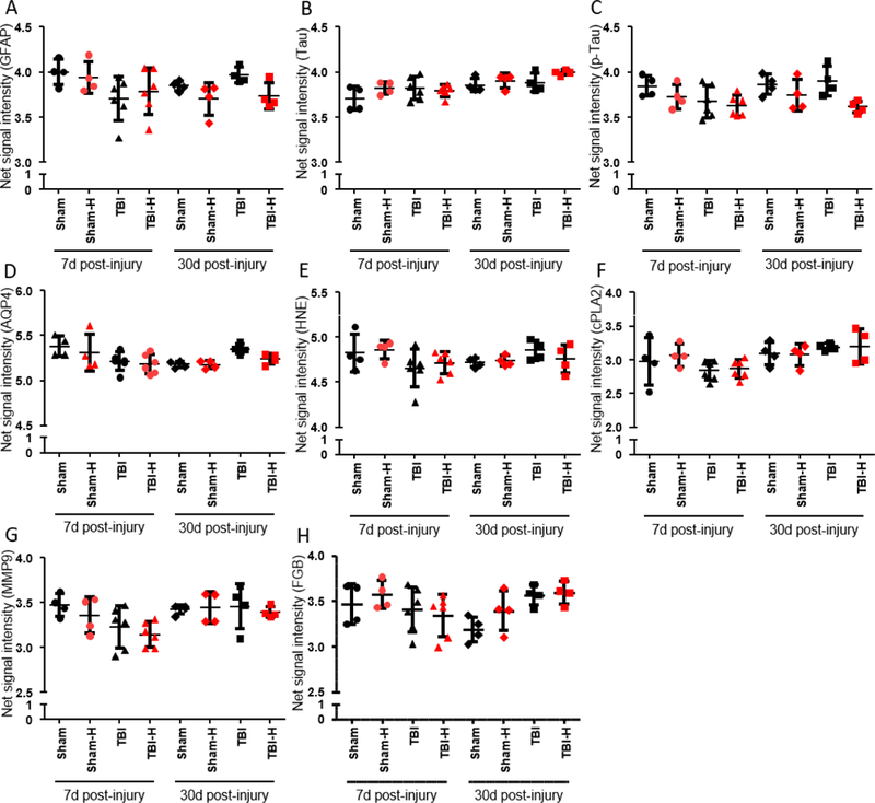
Fig. 5.

Plasma levels of select proteins measured in duplicate low-hemolysis (black) vs. high-hemolysis (red) samples collected at 7 and 30 days post-TBI or sham. Hemolysis did not appear to increase the levels of any tested marker. However, the hemoglobin content of high-hemolysis plasma samples may have interfered with protein measurements, resulting in lower values in high-hemolysis samples relative to their low-hemolysis counterparts. (A) Glial fibrillary acidic protein (GFAP) is an intermediate filament with diverse functions in the CNS, such as cytostructural integrity. (B) Tau is associated with microtubules in the nervous system. (C) Hyperphosphorylation of Tau (pTau) results in the protein’s dissociation from microtubules, compromising structure and, in turn, function. P-Tau values were significantly lower in one high-hemolysis sample (p < 0.05, Wilcoxon test). (D) Aquaporin-4 (AQP4) is a water-selective channel protein predominantly found in the brain. Under physiological and pathological conditions, AQP4 plays an important role in water homeostasis. (E) 4-Hydroxynonenal (HNE) is a byproduct of lipid peroxidation, indicative of oxidative stress. (F) Cytosolic phospholipase-A2 (cPLA2) becomes catalytically active in the presence of free Ca2+ as seen in stimulated cells; cPLA2 hydrolyzes cellular phospholipids whose breakdown products contribute to inflammation and degeneration. (G) Matrix metalloproteinase-9 (MMP9) plays an essential role in local proteolysis of the extracellular matrix. (H) Fibrinogen (FGB) was the first hemostatic factor to be described as a target for miRNAs (Fort et al., 2010). FGB levels increase in the acute phase of the injury and in inflammatory states. Data are presented as the mean ± SD. Abbreviations: d, days; H, high hemoglobin samples based on Nanodrop measurements; SD, standard deviation; TBI, traumatic brain injury.
