
Fig. 4.
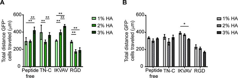
Migratory response of GFP+ and GFP− cells is influenced by %HA and type of peptide. There was an overall decrease in GFP+ and GFP− cell migration on RGD-functionalized hydrogels. While TN-C-functionalized hydrogels had a similar effect on both GFP+ and GFP− cells, IKVAV-functionalized hydrogels had an opposite effect on the migratory response of the two cell types. A) The migratory response of GFP+ cells was significantly affected by %HA within each peptide group. Two-way ANOVA showed significance for peptide/%HA interaction (p < 0.0001), peptide (p < 0.0001), and %HA (p < 0.0001). Tukey’s post hoc test revealed the effect of %HA on the different peptide groups (*p < 0.05, **p < 0.01, ***p < 0.001). B) The migratory response of GFP− cells was less affected by %HA and peptide type. Two-way ANOVA showed significance for %HA (p < 0.05) and type of peptide (p < 0.0001). Tukey’s post hoc test revealed difference in cell migration by IKVAV-functionalized hydrogels between 1% and 3% HA (*p < 0.05). GFP+ and GFP− cells were imaged and tracked every hour for 12 h. Error bars = SD; n ≥ 100 cells per %HA-peptide combination. Three independent experiments were averaged with n ≥ 3 technical replicates per experiment.