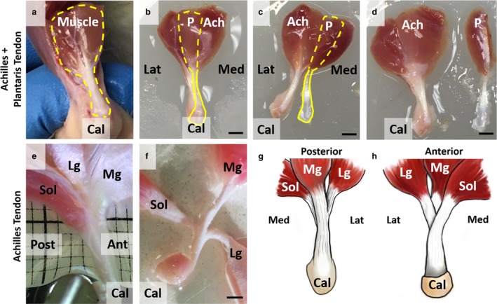
Figure 1.

Gross morphology of plantaris and Achilles tendons. (a) Plantaris (P) and Achilles (Ach) tendons are bound together in situ (dashed yellow line). (b) Plantaris muscle is posterior to Achilles muscle complex (dashed yellow line), and plantaris tendon is visible above Achilles tendon (solid yellow line). (c) By cutting connective tissue binding plantaris and Achilles tendons from the insertion, these two tendons can be completely separated (d). (e) Achilles tendon was further dissected and is a fusion of three tendons originating from soleus (Sol), lateral gastrocnemius (Lg), and medial gastrocnemius (Mg) muscles. (f) Sub‐tendons are tightly fused, and further separation of tendons is difficult without directly cutting the tendon. Scale bars: 2 mm. We created schematics of (g) posterior and (h) anterior views to show the complex structure of Achilles tendon.
