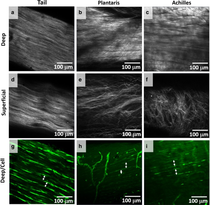
Figure 4.

Collagen alignment and cell morphology. (a‐c) Collagen alignment was viewed with SHG signals. Collagen was aligned at deep planes, away from the skin, for tail, plantaris, and Achilles tendons. (d‐f) Whereas the collagen alignment was not different between the superficial, close to the skin, and deep planes for the tail tendon, plantaris and Achilles tendons had an additional layer of a complex meshwork of collagen or peritenon. (g,h) In deep planes, elongated cells outlined collagen bundle (white arrows), which matched the sizes of fiber observed with histology. Thus, we define fiber as a bundle of collagen with diameter 10–50 μm outlined by elongated cells. No fascicle boundary was observed.
