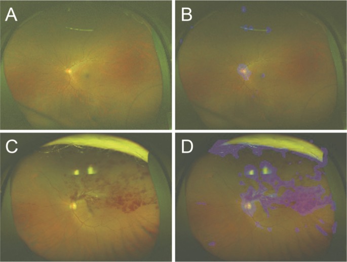
Figure 4. Representative ultrawide-field fundus images and their corresponding heat maps.

The ultrawide-field fundus image without BRVO (A) and its corresponding heat map (B); with BRVO (C) and its corresponding heat map (D). In the image without BRVO, the deep convolution neural network focused on the optic disc (B; blue color). Meanwhile, in the image with BRVO, the deep convolution neural network focused on the optic disc and retinal hemorrhages (D; blue color).
