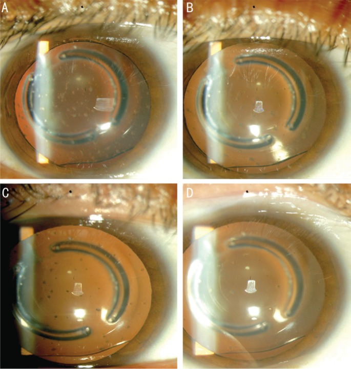
Figure 2. Consecutive clinical retro-illumination slit-lamp photographs of the left eye.

A: The white deposits were numerous during the first day of diagnosis; B, C, D: After only intense topical steroid treatment the deposits decreased over time until the ICL almost become clear.
