
Fig. 5.
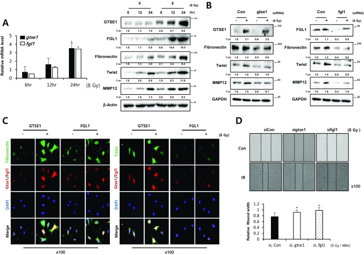
Radiation induces EMT through gtse1 or fgl1. a Expression of GTSE1 and FGL1 in L132 cells was examined by qRT-PCR (left) and Western blotting (right) analysis after 8Gy radiation (IR) at indicated time points. b Western blot analysis in L132 cells after transfection of siRNA gtse1 or fgl1 with or without 8Gy radiation (IR). The fold increase of each time point mRNA was normalized with the non-irradiated cells (n = 3, mean ± SD, *p < 0.05 vs. corresponding control, paired sample t test) and protein levels were quantified using Image J software, and data are expressed as the fold change relative to the negative control. c Co-immunofluorescence staining of Fibronectin or Twist (green) and GTSE1 (red) or FGL1 (red) after 24 h of irradiation (8 Gy) in A549 cells. Nuclei were counterstained with DAPI (blue). d Wound healing assays in A549 cells after transfection of siRNA gtse1 or fgl1 with or without irradiation (8 Gy). Cell movement into wound was shown at 48h post-scratch. Graphs represent relative wound width (mean ± SD, *p < 0.05 vs. corresponding control, paired sample t test). All representative photomicrographs magnification, × 100; bar, 50 μm