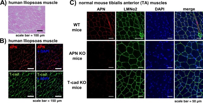
Figure 1.
Localization of APN and T-cad in intact human and mouse muscle. A series of paraffin sections of human iliopsoas muscle autopsy specimen was stained with H&E (A), with anti-human APN and with anti-human T-cad (B). Cell nuclei were counterstained with DAPI. Scale bars, 100 μm for (A), 150 μm for (B). (C) Confocal immunofluorescence micrographs of tibialis anterior (TA) muscle of WT, adiponectin KO (APN KO), and T-cadherin KO (T-cad KO) mice. Muscle tissues were stained with anti-adiponectin (APN; red), anti-laminin α2 (LMNα2; green). Cell nuclei were counterstained with DAPI (blue). Scale bars, 50 μm.

