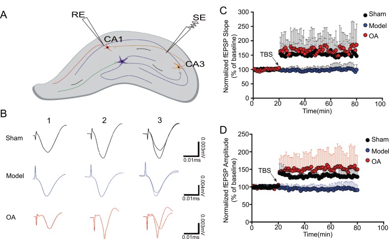
Fig. (4).
OA rescues Aβ-induced deficits in long-term potentiation (n=3). (A) Position of stimulation electrode (SE) and recording electrode (RE); (B) 1: potential of resting state; 2: 60 min after TBS; 3: combination of (1) and (2) in each group; (C) slope change in each group. Kruskal-Wallis test showed that the slope rate significantly decreased after high frequency stimulation in the model group compared with that in the sham operation group (P = 0.0001). While compared with model group, the amplitude significantly increased after high frequency stimulation in the OA group (P=0.0001) (D) amplitude change in each group. Kruskal-Wallis test showed that the amplitude significantly decreased after high frequency stimulation in the model group compared with that in the sham operation group (P=0.0001). While compared with model group, the amplitude significantly increased after high frequency stimulation in the OA group (P=0.0001)

