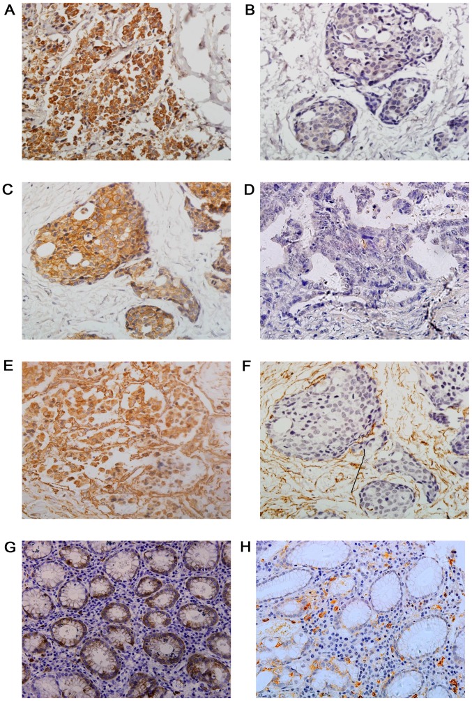
Figure 1.
Immunohistochemical staining of STMN1, E-Cad and VIM in GC and adjacent normal tissues. Representative images displaying (A) positive and (B) negative STMN1 expression, (C) positive and (D) negative E-cad expression, and (E) positive and (F) negative VIM expression in GC tissues, as well as the (G) positive and (H) negative STMN1 expression in adjacent normal tissues. Magnification, ×400. STMN1, Stathmin1; E-Cad, E-cadherin; VIM, vimentin; GC, gastric cancer.

