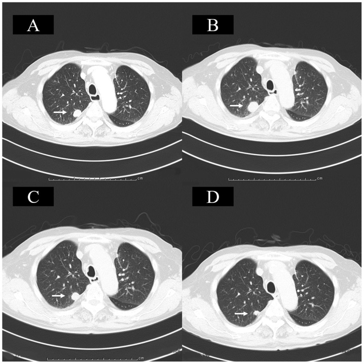
Figure 1.
Chest CT. (A) Chest CT revealed a metastatic lung tumor in the right upper lobe prior to nivolumab therapy. (B) Following the 6th course of nivolumab therapy, CT revealed that the lung metastases had grown, representing progressive disease. (C) Following the 10th course of nivolumab therapy, CT demonstrated that the lung metastases had shrunk to their pretreatment size. (D) Following the 14th course of nivolumab therapy, CT showed that the lung metastases had shrunk when compared with that observed prior to treatment. Arrows indicate the area of lung metastasis. CT, computed tomography.

