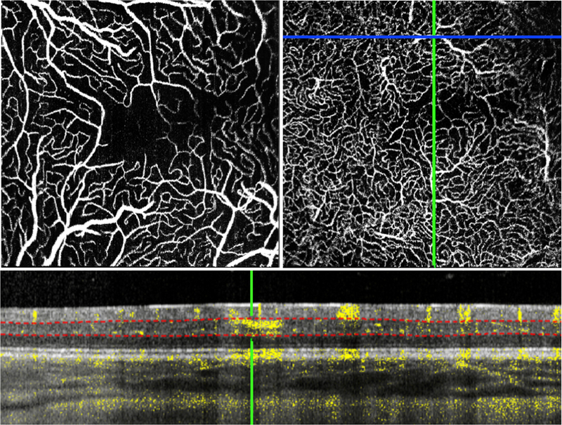
Figure 1.

The macula of left eye of an infant with stage 3 ROP, regressed after bevacizumab treatment, imaged at 73 weeks postmenstrual age showed an irregular angular vessel pattern with several large vessels in the superficial vascular complex (A) diving down into the deeper layers of the retina (examples circled in red in B and C). A B-scan shows one of these diving vessels (red arrow at the green crosshair) penetrating into the inner nuclear layer. Red dotted lines indicate the manually placed segmentation boundaries for the DVC.
