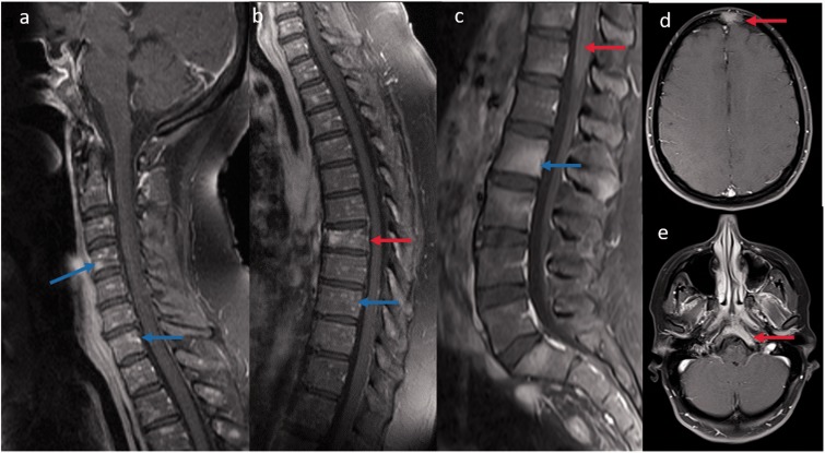
Figure 4.
(a, b) Sagittal T1-weighted post-contrast images reveal diffuse peppered enhancement of the cervical and thoracic vertebrae bone marrow (blue arrow) without any leptomeningeal and cord involvement. Associated T7 vertebral pathological collapse (red arrow). Biopsy revealed non-caseating granuloma. (c) Sagittal T1-weighted post-contrast image in another patient reveals multifocal areas of bone marrow enhancement (blue arrow) along with leptomeningeal enhancement (red arrow). Axial T1-weighted post-contrast brain images of the same patient also showed bony enhancement of frontal and clivus (red arrow) regions.

