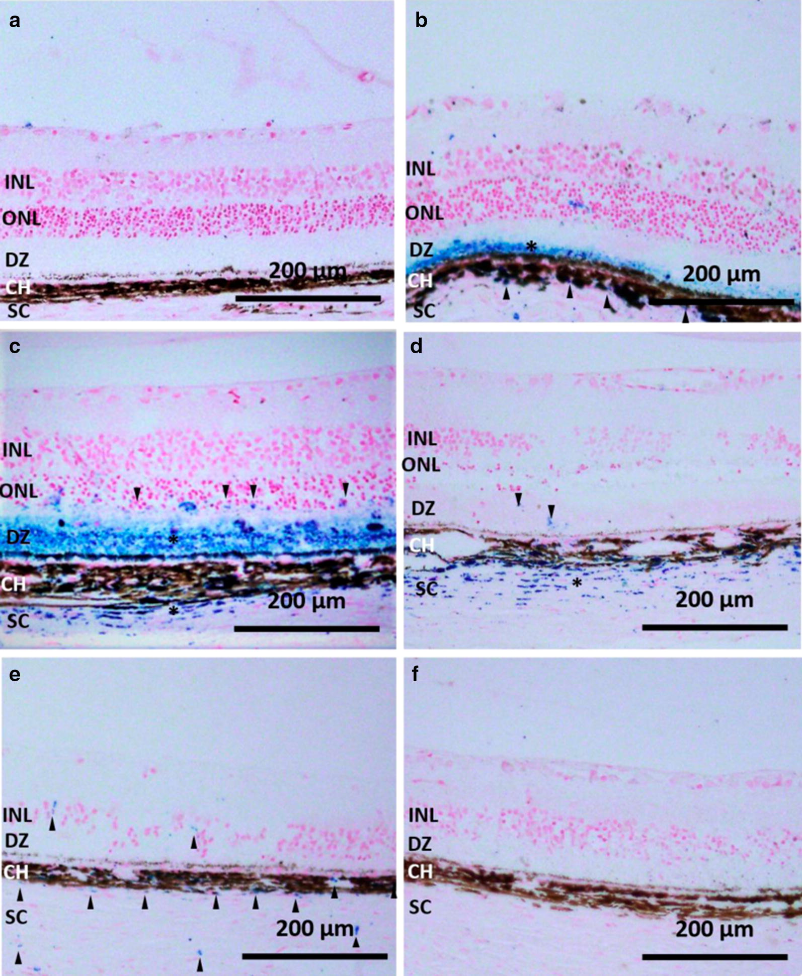
Fig. 2.

IO/HSA NPs localization in the RCS retina. Sections of RCS retinas removed 2 h (b),1 week (c), 4 weeks (d), 6 weeks (e), and 12 weeks (f) following injection as well as the contralateral non injected eye of the same rat shown in panel b (a) were stained with Prussian blue and counter stained with nuclear fast red. Asterisks highlight layers with positive Prussian blue staining, and arrowheads highlight focal Prussian blue staining. Scale bar 200 µm. INL inner nuclear layer, ONL outer nuclear layer, DZ debris zone, CH choroid, SC sclera
