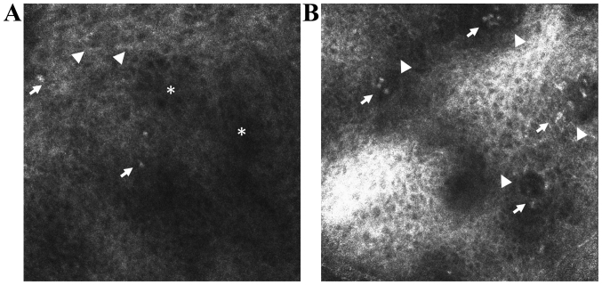
Figure 2.
In vivo RCM features of lichen planus. (A) RCM image (0.5×0.5 mm) at the level of the granular-spinous layer showing increased intercellular spaces (spongiosis) (*), large, polygonal cells (hypergranulosis in a wedge-shaped pattern that corresponds to the Wickham's striae) (▶) and inflammatory cells that appear as roundish bright structures (→). (B) RCM image (0.5×0.5 mm) at the level of the epidermal-dermal junction showing non-edged and non-rimmed dermal papillae (▶) due to inflammatory cell infiltrate (→).

