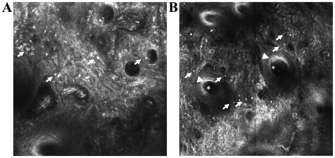
Figure 3.
In vivo RCM features of discoid lupus erythematosus. (A) RCM mosaic (1×1 mm) at the epidermal-dermal junction level showing disappearance of the papillary rings due to infiltrates of inflammatory cells (→). (B) RCM mosaic (1×1 mm) showing dillated follicle (*) with infundibular hyperkeratosis (▶) surrounded by inflammatory cells (→).

