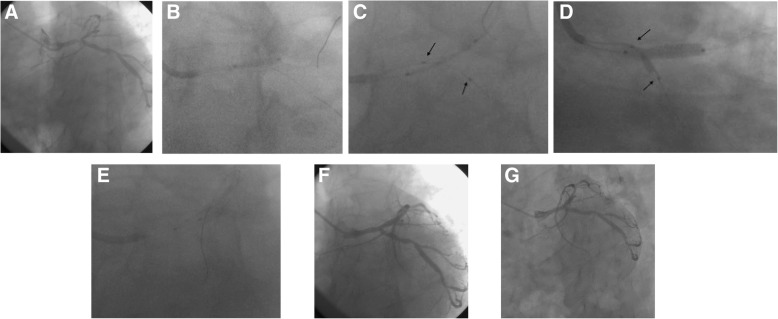
Fig. 2.
a Lesion angiography before procedure; (b) The MV lesion was managed with a standard semi-compliant balloon predilatation; (c)A stent with adequate size and length was used to cover the MV lesion, then a fitful balloon was sent into the SB; the proximal markers of the SB balloon were not beyond that of the MV stent(Black Arrow), and the distal markers covered the SB ostium lesion(Black Arrow); (d)The MV stent balloon and SB balloon(Black Arrow)were inflated simultaneously; the SB balloon was inflated to the normal pressure; (e) For optimization of MV stent apposition, the proximal optimization technique (POT) was performed with a short non-compliant balloon; (f)Immediate angiography after operation;(g) 9 months later angiography after operation

