Fig. 3.
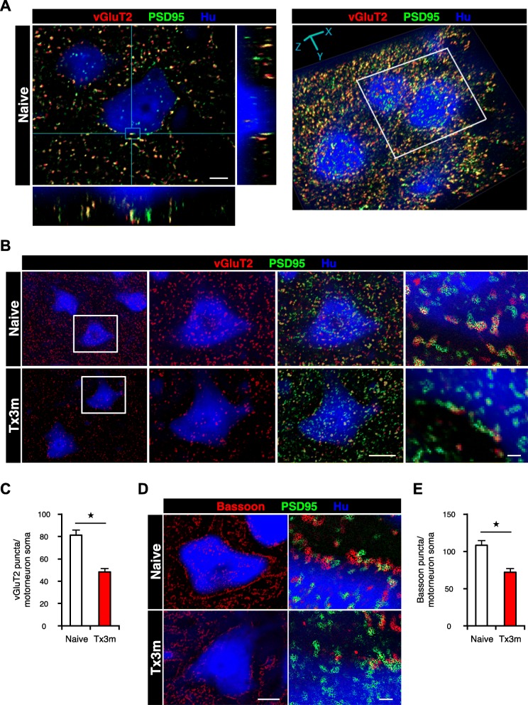
The presynaptic input to the lumbar motor neurons decreases in the chronic phase of SCI. a Representative images of lumbar motor neurons, showing 3D colocalization of vGluT2-positive presynaptic boutons (red) and PSD95-positive postsynaptic boutons (green). b Immunohistochemical analysis of excitatory synaptic boutons in lumbar motor neurons, stained for vGluT2 (red), PSD95 (green), and Hu (blue). The two middle images are magnifications of the boxed areas. c Quantification of the vGluT2-positive excitatory presynaptic boutons in lumbar motor neurons of the Naive and Tx3m groups (n = 8 neurons from 4 mice). d Immunohistochemistry for pan-synaptic boutons in lumbar motor neurons, stained for Bassoon (red), PSD95 (green), and Hu (blue). e Quantification of Bassoon-positive pan-presynaptic boutons in lumbar motor neurons of the Naive and Tx3m groups (n = 8 neurons from 4 mice). *P < 0.05, Wilcoxon rank sum test (c and e). Data are presented as the mean ± SEM. Scale bars: 5 μm (a); 10 μm (b, two middle panels; d, left panel); 1 μm (b and d, right panels)

