Fig. 6.
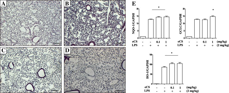
The effect of intratracheal delivery of eCS on lung inflammation and the expression of Nrf2-dependent genes in an LPS-induced ALI mouse model. C57BL/6 mice (n = 5/group) received sham (a) or a single, 2 mg/kg body weight of i.t. LPS (b, c, and d). At 2 h after LPS treatment, mice received a single, 0.1 mg/kg body weight of i.t. eCS (c) or 1 mg/kg body weight of i.t. eCS (d). At 24 h after LPS administration, the lungs of mice were harvested and stained with HE for histological examination. Data are representatives of at least five different areas of a lung (bar, 200× magnifications). e Total RNA extracted from the harvested lungs (n = 5/group) was analyzed by semi-quantitative RT-PCR to assess expressions of NQO-1, HO-1, and GCLC. The intensity of each PCR band was measured by densitometric analysis (ImageJ) and normalized to GAPDH intensity. * P was less than 0.05, compared with the LPS treated (post-ANOVA comparison with Tukey’s post hoc test)

