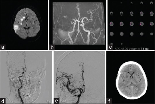
Figure 1.

A 69-year-old female with right middle cerebral artery territory stroke. (a) Diffusion-weighted images show the right middle cerebral artery territory infarcts. (b) Time-of-flight magnetic resonance angiogram demonstrates right middle cerebral artery occlusion. (c) Rapid processing of perfusion and diffusion images indicating ischemic core (apparent diffusion coefficient <620) of 33 ml. (d) Preprocedure digital subtraction angiogram showing the right middle cerebral artery occlusion. (e) Postthrombectomy digital subtraction angiogram image showing recanalization of the middle cerebral artery branches. (f) Computed tomography brain done after 24 h shows small infarct corresponding to the core volume
