Figure 1.
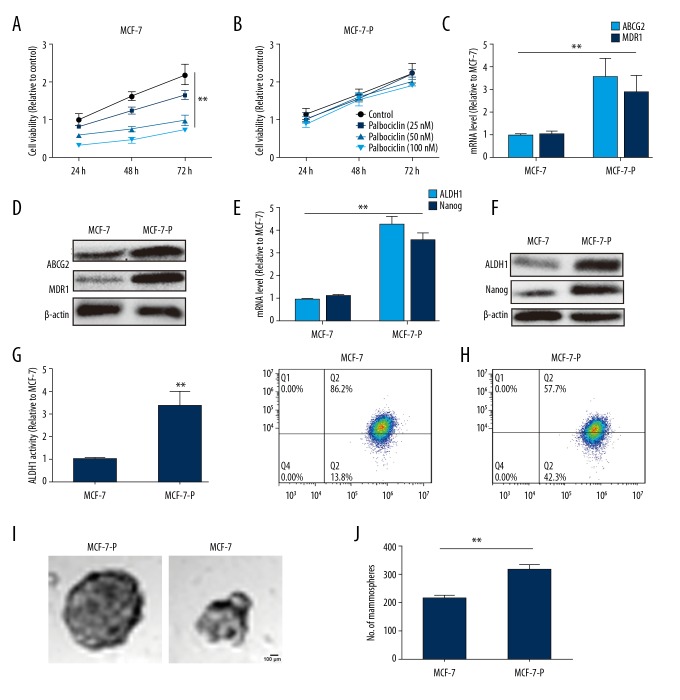
MCF-7-P cells exhibit palbociclib resistance and higher stemness. (A) MCF-7 cells were treated with different concentration of palbociclib, and after 24, 48, and 72 hours, the cell viability was analyzed by MTT assay. (B) MCF-7-P cells were treated with different concentration of palbociclib, and after 24, 48, and 72 hours, the cell viability was analyzed by MTT assay. (C) mRNA level of drug resistance-related proteins ABCG2 and MDR1 was detected in MCF-7 and MCF-7-P cells. (D) Protein levels of ABCG2 and MDR1 was examined in MCF-7 and MCF-7-P cells. (E, F) mRNA and protein levels of stemness markers ALDH1 and Nanog were determined in MCF-7 and MCF-7-P cells. (G) ALDH1 activity was measured in MCF-7 and MCF-7-P cells. (H) The CD44+/CD24- cell sub-population was detected in MCF-7 and MCF-7-P cells. (I, J) The cells spheroid formation ability was evaluated in MCF-7 and MCF-7-P cells via measuring the spheroids size and number. Data were presented as mean ± standard deviation; ** P<0.01 versus MCF-7.

