Figure 4.
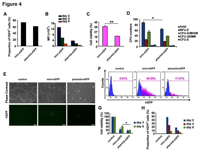
BF minicircle vector electroporation in CD34+ HSCs and hESCs. (A) Proportion of eGFP-positive cells in CD34+ HSCs transfected with BF minicircle vector and plasmid encoding eGFP 48 hours after electroporation. (B) The MFI of eGFP-positive cells in days 2, 4 and 6 after mini-eGFP and plasmid-eGFP electroporation. (C) Cell viability of CD34+ HSCs transfected with BF minicircle vector and plasmid encoding eGFP 48 hours after electroporation. (D) Quantification of total CFU numbers and the numbers of BFU-E, CFU-G/M/GM, CFU-GEMM and CFU-E of CD34+ HSCs transfected with BF minicircle vector and plasmid encoding eGFP. (E) Typical images of H9 hESCs transfected with BF minicircle vector or plasmid encoding eGFP, 48 hours after electroporation. Scale bar=100 µm. (F) FACS analysis of eGFP-positive cells in H9 transfected with BF minicircle vector or plasmid encoding eGFP, 48 hours after electroporation. (G) Cell viability of H9 transfected with BF minicircle vector or plasmid encoding eGFP, 2 days and 6 days after electroporation. (H) Proportion of eGFP-positive cells in H9 transfected with BF minicircle vector or plasmid encoding eGFP, 2, 4 and 6 days after electroporation. (A–D) Data of CD34+ HSCs from one donor and more than three donor samples were detected. Unpaired multiple two-tailed t-test. The asterisks indicate significant differences between the mini-eGFP group and the plasmid-eGFP group: *p<0.05, **p<0.01. BF, bacteria-free; CFU, colony forming unit; eGFP, enhanced green fluorescent protein; FACS, fluorescence-activated cell sorting; HSC, haematopoietic stem cell; MFI, mean fluorescence intensity.

