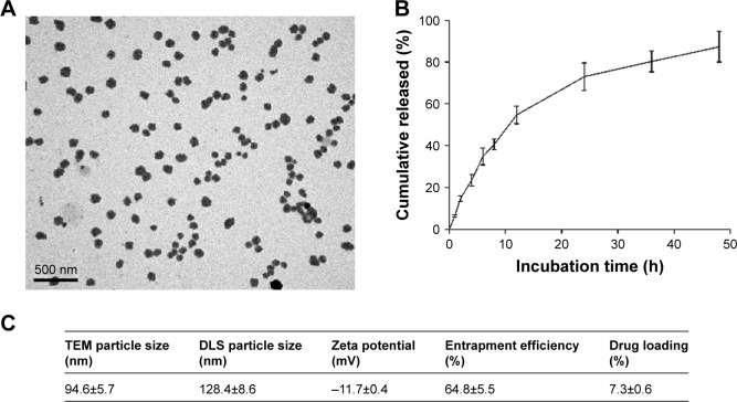
Figure 1.
Characterization of PLGA-CPT NPs.
Notes: (A) Representative transmission electron micrograph of NPs. (B) The in vitro release of CPT from PLGA-CPT NPs into buffer. (C) Physicochemical characterization of PLGA-CPT NPs. Data expressed as mean±SD from three independent experiments.
Abbreviations: DLS, dynamic light scattering; NP, nanoparticle; PLGA-CPT, camptothecin-encapsulated poly(lactic-co-glycolic acid); TEM, transmission electron microscopy.

