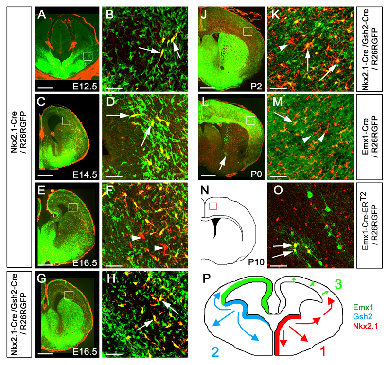
Figure 1.
Three successive waves of OLs generated from distinct precursor populations at different times during forebrain development. (a, b) Generation of the first wave of Pdgfra+ OLPs begins at E12.5 from Nkx2.1-expressing precursors in the MGE/AEP. These OLPs express GFP (green) in Nkx2.1-Cre/ Rosa26R-GFP embryos (arrows in b). (c, d) By E14.5 Pdgfra+, GFP+ cells start to appear at the cortico-striatal boundary in Nkx2.1-Cre/ Rosa26R-GFP embryos (arrows in d), demonstrating migration of MGE/AEP-derived OLPs into the developing cortex. (e, f) By E16.5 a new wave of Pdgfra+, GFP-negative OLPs is observed in Nkx2.1-Cre/ R26R-GFP cortex; these must be generated from Nkx2.1-negative precursors (arrowheads in f). (g, h) All Pdgfra+ cells in the telencephalon co-express GFP in Nkx2.1-Cre/ Gsh2-Cre/ Rosa26R-GFP triple-transgenic embryos (arrows in h), indicating that all OLPs are derived from the ventral forebrain (MGE/AEP + LGE/CGE) at this stage. (i, j) Another wave of Pdgfra+, GFP-negative OLPs appears in the telencephalon of early postnatal Nkx2.1-Cre/ Gsh2-Cre/ Rosa26R-GFP mice (arrowheads in j). (k, l) Emx1-expressing cortical precursors begin to generate OLPs/OLs after birth (arrows in l). (m, n) Tamoxifen administered once to pregnant Emx1-CreERT2 females at E9.5, before invasion of the cortex by ventrally-derived OLPs, activates Cre recombination in the embryos and results in expression of the GFP reporter in a subset of Sox10+ OLPs (arrows in n)- which must therefore have been generated from endogenous cortical precursors. (o) Conclusion: three sequential waves of OLPs are generated from different parts of the telencephalic VZ: 1) from Nkx2.1-expressing precursors starting at E12.5, 2) from Gsh2-expressing LGE/CGE precursors starting at E15.5 and 3) from Emx1-expressing cortical precursors starting around birth (P0). Scale bars: (a, c, e, g, i, k) 500 μm; (b, d, f, h, j, l, n) 60 μm.

