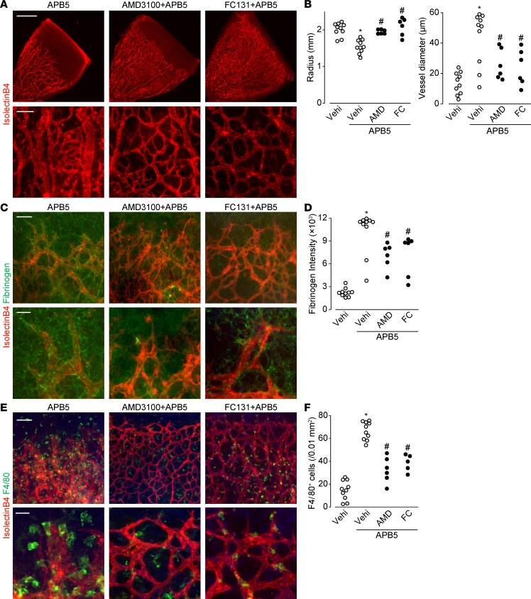
Figure 3. CXCR4 inhibition abrogated the progression of APB5-induced BRB breakdown and macrophage infiltration.
(A) Representative images of retina immunostaining of isolectin B4 (red) on P8. Scale bar: 500 μm (top); 50 μm (bottom). (B) The effect of AMD3100 (AMD) or FC131 (FC) on APB5-treated retinal radius and vessel diameter on P8 (n = 6–10). (C) Representative images of retina immunostaining of isolectin B4 (red) and fibrinogen (green) on P8. Scale bar: 100 μm (top); 30 μm (bottom). (D) The effect of AMD3100 or FC131 on APB5-treated retinal fibrinogen intensity on P8 (n = 6–10). (E) Representative images of retina immunostaining of isolectin B4 (red) and F4/80 (green) on P8. Scale bar: 100 μm (top); 30 μm (bottom). (F) The effect of AMD3100 or FC131 on APB5-treated retinal F4/80-positive cells on P8 (n = 6–10). Significantly different from the results in PBS- or APB5-treated mice at *P < 0.05, significantly different from the results in PBS-treated mice. #P < 0.05, significantly different from the results in APB5-treated mice.

