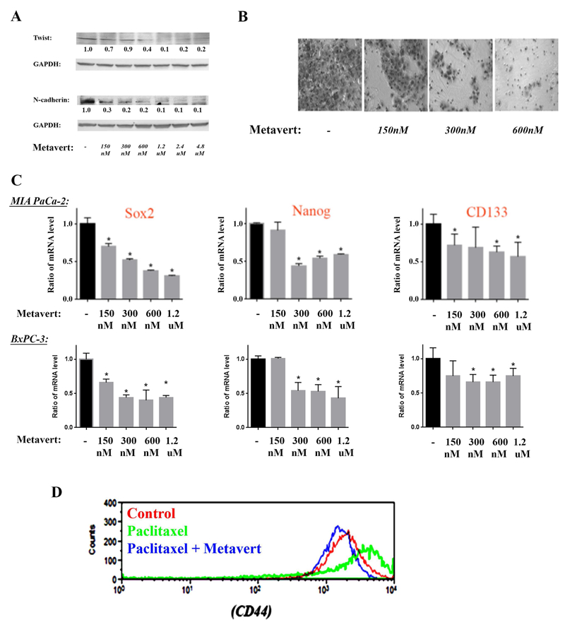
Figure 3: Metavert prevents migration, EMT and cancer stemness markers in cancer cells.
MIA PaCa-2 and BxPC-3 cells were cultured for 72h with different doses of metavert. (A) Protein levels in MIA PaCa-2 were measured by Western. Blots were re-probed for GAPDH to confirm equal loading. (B) MIA PaCa-2 cell migration was measured by Matrigel Invasion Assay. After 72h treatment, 100,000 cells were re-plated overnight for the invasion assay. The total number of cells did not change during the overnight invasion assay. (C) mRNA levels were measured by RT-PCR in MIA PaCa-2 and BxPC-3 cells. (D) Level of CD44 was measured by flow cytometry using CD44 (PE/Cy7) antibody in MIA PaCa-2 cells treated with metavert (600nM) or Paclitaxel (10nM).

