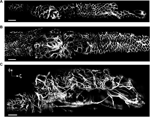
Figure 4:

Vascular changes during intestinal tumorigenesis.
Cancer growth was induced by administration of adenoviral Cre into the bowel epithelia of Apc mice to inactivate the Apc gene. (A–C) After making a midline incision, the jejunum was exteriorized outside the abdomen, and opened with a 18 gauge needle. A side-view endoscope was inserted into the bowel. Fluorescence image of colorectal vasculature in a floxed Apc mouse at 11 weeks (A) and 13 weeks (B) after administration of the carcinogenetic vector, and in a large lesion at week 17 (C) in another Cre-treated mouse. Blood vessels are visualized by fluorescence after intravenous injection of FITC-dextran, and show a progressive disorganization in the course of carcinogenesis. Scale bars, 200 μm. (Reproduced by permission from Macmillan Publishers Ltd: Kim P et al. Nat Methods, Copyright 2010).
