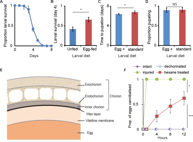
Fig 1. Wax layer of the eggshell prevents egg cannibalism.
(A) Survival period of freshly hatched larvae held individually without food on agar (mean = 3.4 ± 0.1 days (SE); n = 4 replicate plates, 24 freshly hatched larvae assayed per replicate), demonstrating the surplus nutrition provisioned within an egg for embryonic development well beyond hatching. (B) Proportion of freshly hatched larvae surviving until day four, in absence of food (unfed) and when allowed to feed on two injured eggs (n = 4 replicate plates per treatment, 24 freshly hatched larvae assayed per replicate). Egg-fed larvae had a higher survival than unfed larvae (mean ± SD). (C, D) Larval developmental period (mean ± SD) and larval survival to pupation (mean ± SD), respectively, when reared on standard diet laced with (blue bars) or without (red bars) extract from crushed eggs (n = 4 bottles/diet, egg density: 200 eggs/bottle). On supplemented diet, larvae developed faster (ANOVA: F1,9 = 9.75, p = 0.014) but survived to a similar extent (ANOVA: F1,9 = 0.07, p = 0.8). (E) Graphical depiction of the egg shell morphology. The outer three layers—the exochorion, the endochorion, and the inner chorion layer—together form the chorion; the thin wax layer (in black) lies between the chorion and the innermost vitelline membrane that envelops the egg. (F) Proportion (mean ± SE) of intact, injured, dechorinated, and hexane-treated eggs cannibalized when confined in agar vials with second-instar larvae over 12 h (n = 10 vials/treatment, with 5 eggs and 10 larvae per vial). Larvae cannibalized hexane-treated eggs but not dechorinated eggs (ANOVA: F1,18 = 21.85, p = 0.0002), however, not to the extent of injured eggs (ANOVA: F1,18 = 7.57, p = 0.0132). Also see S1 Fig. ***p < 0.001, *p < 0.05. Data underlying this figure can be found in S1 Data. NS, not significant

