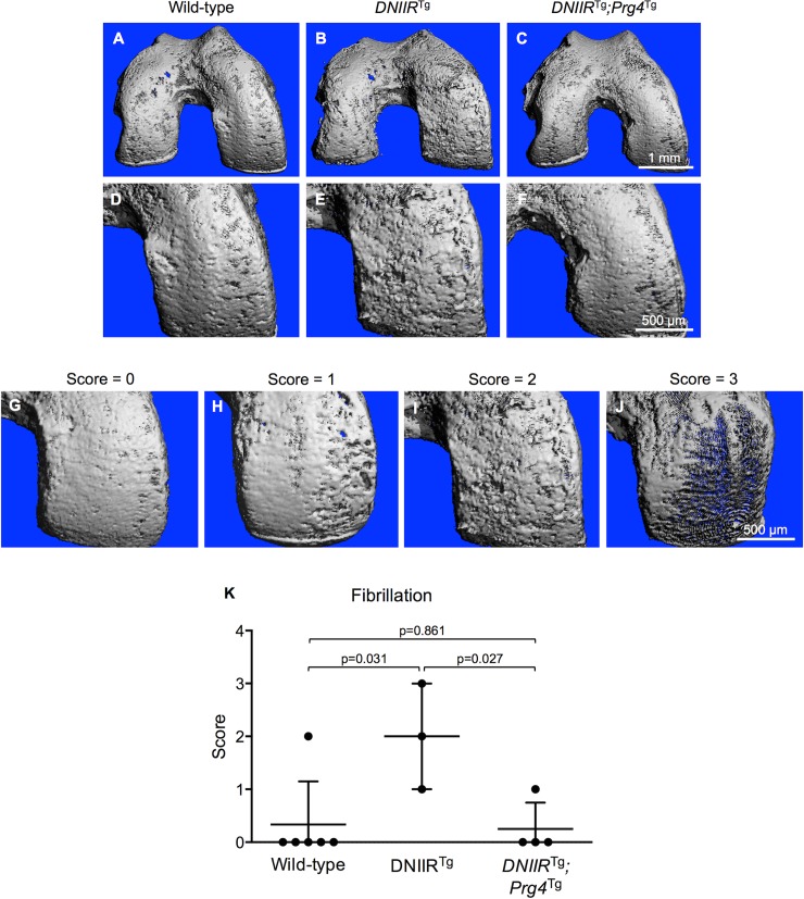
Fig 3. Prg4 prevents fibrillation of articular cartilage in DNIIRTg mice.
μCT imaging was used to obtain 3D images of the surface of femoral articular cartilage in 18-month-old mice. (A-B, D-E) Wild-type mice exhibited mostly smooth cartilage, while DNIIRTg mice exhibited more fibrillation, especially in load-bearing regions. (C and F) DNIIRTg;Prg4Tg mice exhibited smoother cartilage than DNIIRTg mice. (D and F) Wild-type cartilage and DNIIRTg;Prg4Tg cartilage appeared similarly smooth. The medial condyle is shown up-close in D-F. To quantify the fibrillation on the medial condyle, we developed a scoring system. (G) A score of 0 corresponds to no fibrillation or very minimal fibrillation; (H) a score of 1 corresponds to fibrillation that partially covers the medial condyle; (I) a score of 2 corresponds to fibrillation that fully covers the medial condyle and consists of mostly shallow fissures; (J) and a score of 3 corresponds to fibrillation that fully covers the medial condyle and consists of mostly deep fissures. Images were scored by an observer blinded to genotype. The following results are reported as “mean (lower bound of the confidence interval, upper bound of the confidence interval).” (K) The fibrillation score was 0.3 (-0.5, 1.2) for wild-type mice (n = 6; 2 female, 4 male), 2.0 (-0.5, 4.5) for DNIIRTg mice (n = 3; 1 female, 2 male), and 0.3 (-0.5, 1.0) for DNIIRTg;Prg4Tg mice (n = 4; all 4 male). There were the following statistically significant differences (reported as “mean difference (lower bound of confidence interval, upper bound of confidence interval)”): wild-type and DNIIRTg mice (-1.7 (-2.9, -0.4)) and DNIIRTg and DNIIRTg;Prg4Tg mice (1.8 (0.4, 3.1)).

