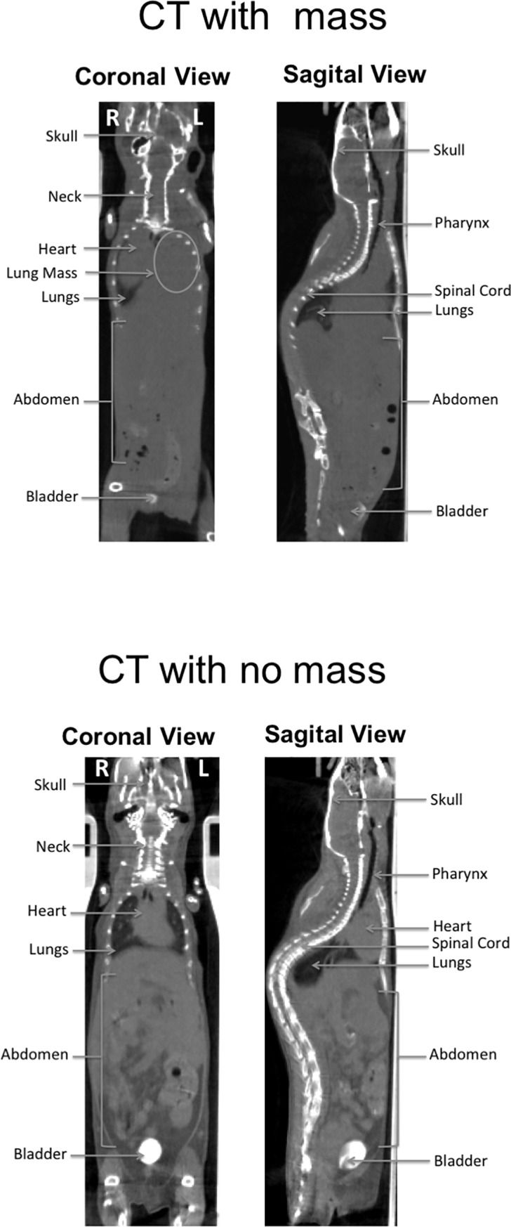
Fig 3. Full body CT scans three weeks post-boost vaccination.

Aged (24–26 months) BALB/c mice were immunized twice with H1-VLP, split-virion vaccine or naïve. Three weeks post-boost, 4–15 mice/group underwent full body CT scans. A) Depicts a mouse with the presence of mass on the left side of its chest. B) demonstrates a relatively healthy aged mouse with no mass detected.
