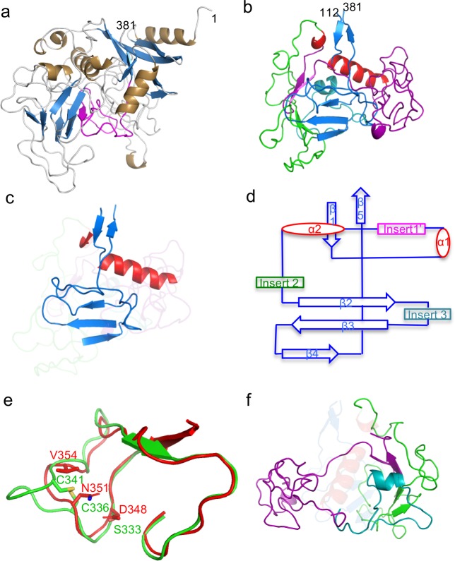
Fig 2. Structure of TaqVP.
a. TaqVP in ribbon representation (α-helices gold, β-strands blue, loops grey, and VR purple). The amino acid positions of the N- and C-termini of TaqVP are indicated. b. TaqVP in ribbon representation with the core elements of the CLec-fold in red (α-helices) and blue (β-strands). Insert 1’, amino acids 129–217, is magenta; insert 2, amino acids 222–296, green; insert 3, amino acids 301–331, teal. c. The core elements of the CLec-fold in TaqVP in ribbon representation (α-helices red, β-strands and loops blue). The inserts are ghosted. d. Topology diagram of the CLec-fold in TaqVP. e. Superposition of a portion of the TaqVP VR (red) and the catalytic site of hFGE (green) in Cα representation. The catalytic triad of hFGE is in bonds representation, as are structurally equivalent or near-equivalent amino acids of TaqVP. f. Inserts of TaqVP in ribbon representation, with core elements of the CLec-fold are ghosted (color coding same as in panel b).

