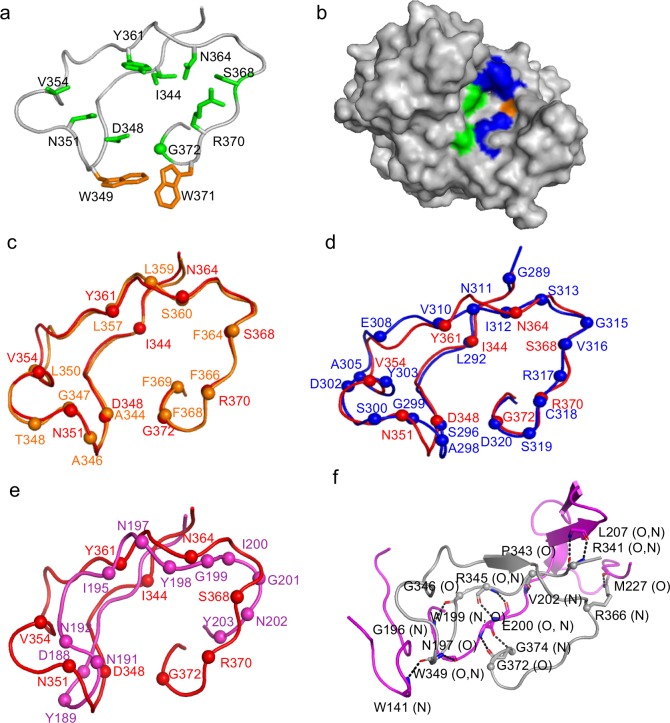
Fig 3. Variable region of TaqVP.
a. VR of TaqVP in ribbon representation. The main chain is in gray, side chains of variable amino acids are in green (sphere is glycine), and nonvariable aromatic amino acids are in orange. b. Surface representation of TaqVP, with the VR facing the viewer. Variable hydrophobic amino acids (I, V, and Y) are green, variable hydrophilic amino acids (S, N, D, and R) blue, and variable glycine pale orange. c. Superposition of the VR of TaqVP (red) and Mtd-P1 (orange) in Cα representation. The spheres represent the position of variable amino acids in each protein. d. Superposition of the VR of TaqVP (red) and TvpA (blue) in Cα representation. e. Superposition of the VR of TaqVP (red) and AvpA (magenta) in Cα representation. f. Stabilization of the main chain of VR (gray, Cα of variable amino acids indicated by spheres) by insert 1’ (magenta) in Cα representation. Dashed line indicates hydrogen bonds.

