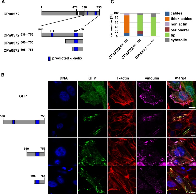
Fig 7. The C-terminus of CPn0572 contains a vinculin-binding domain.
(A) Schematic representation of CPn0572 with its C-terminal variants. ABD-C, black box; ABD, white box; predicted α-helices, dark blue boxes (labeled H1 and H2). Numbers indicate amino acid positions. (B) Confocal fluorescence microscopy analysis of transfected cells expressing GFP or GFP-CPn0572 and vinculin fused to SNAP. HEp-2 cells were transfected for 18 h before fixation. F-actin was visualized with phalloidin (red), vinculin with SNAP-cell SiR647 (magenta) and DNA with DAPI (blue). Bars: 10 μm. (C) Quantification of localization phenotypes of GFP-CPn0572 variants shown in (B). n ≥ 90 cells per transfected plasmid. All quantifications were reproducible and analyzed from triplicates.

