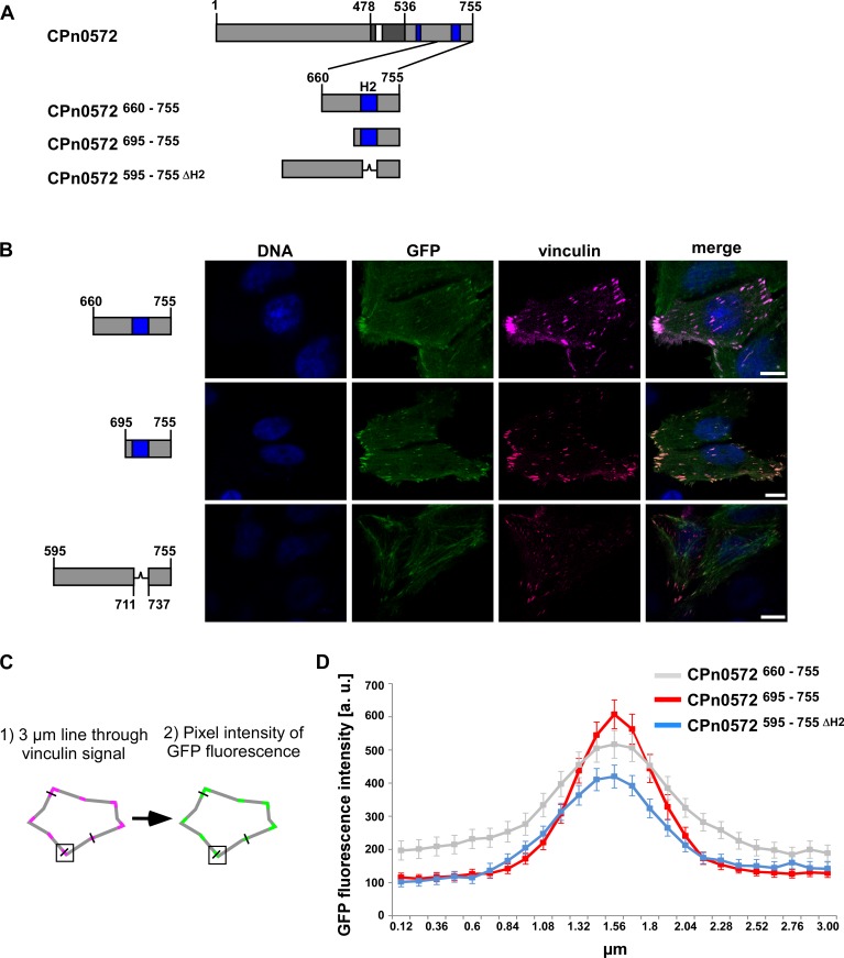
Fig 8. Helix 2 of CPn0572 plays a role in vinculin association.
(A) Schematic representation of CPn0572 and variants thereof. (B) Confocal fluorescence microscopy analysis of transfected cells expressing CPn0572 variants fused to GFP and vinculin fused to SNAP. HEp-2 cells were transfected for 18 h before fixation. Vinculin was visualized with SNAP-cell SiR647 (magenta) and DNA with DAPI (blue). Bars: 10 μm. (C) Schematic representation of the methodology used for the quantification of GFP fluorescence intensity colocalized with vinculin. (D) GFP fluorescence intensity of GFP-CPn0572 variants that colocalize with vinculin puncta. arbitrary units (a.u.). n = 90 independent measurements for each construct analyzed from triplicates.

