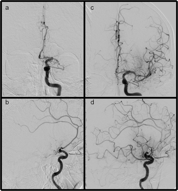
Fig 1.
Ap and lateral views of LAO and eTICI2b50 (mTICI2b) reperfusion results (a+b) ap and lateral views showing a M1 occlusion of the left side (c+d) after recanalization the inferior MCA trunk remains occluded, additionally the frontal division is not fully reperfused. Severe filling defects can be seen in the parietal and frontal part of the MCA territory.

