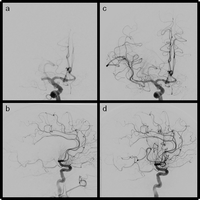
Fig 2.
Ap and lateral views of LAO and eTICI2b67(mTICI2b) reperfusion results (a+b) ap and lateral views of a M1 occlusion of the right side (c+d) ap and lateral view after reperfusion showing a remaining MCA M2 division being still occluded which is led to incomplete reperfusion (central and parietal MCA territory not fully reperfused).

