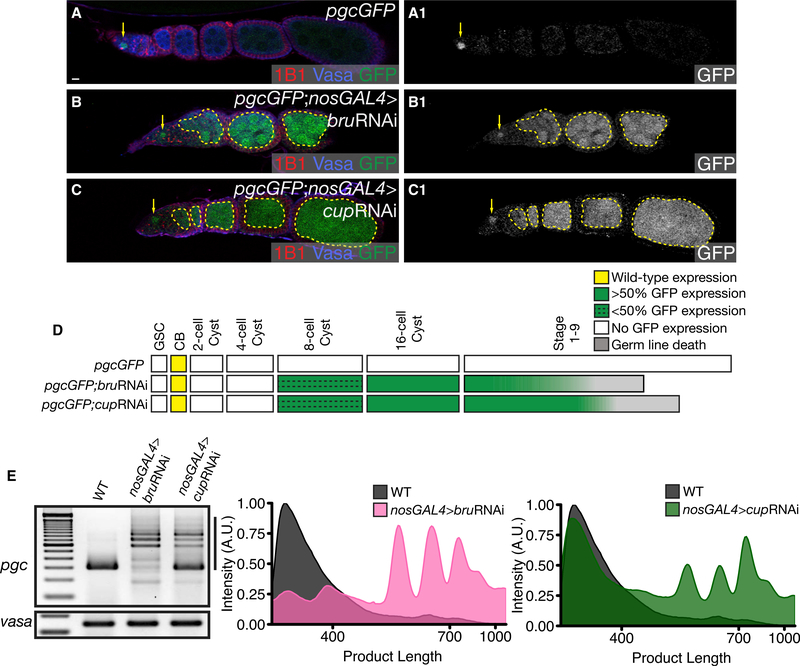
Figure 6. Bru and Cup Regulate Pgc Translation in the Later Stages of Oogenesis.
(A) An ovariole of a pgcGFP ovary stained with 1B1 (red), Vasa (blue), and GFP (green) shows expression of GFP in the pre-CB (arrow).
(B) An ovariole of a pgcGFP; nosGAL4>bruRNAi ovary stained with 1B1 (red), Vasa (blue), and GFP (green) aberrant expression of GFP beyond the 16-cell cyst (12% from 8-cell cyst onward, 100% from 16-cell cyst onward, n = 25) (dashed outline).
(C) An ovariole of a pgcGFP; nosGAL4>cupRNAi ovary stained with 1B1 (red), Vasa (blue), and GFP (green) shows aberrant expression of GFP from the later cyst stages (20% from 8-cell cyst onward, 100% from 16-cell cyst onward, n = 30) (dashed outline). The GFP channel is shown in A1–C1.
(D) A developmental profile of GFP expression when Bru and Cup are depleted in the germline.
(E) PAT assay analysis of pgc poly(A)-tail length of pgc RNA when Bru and Cup are depleted in the germline.
Scale bars, 10 μm. See also Figure S6.

